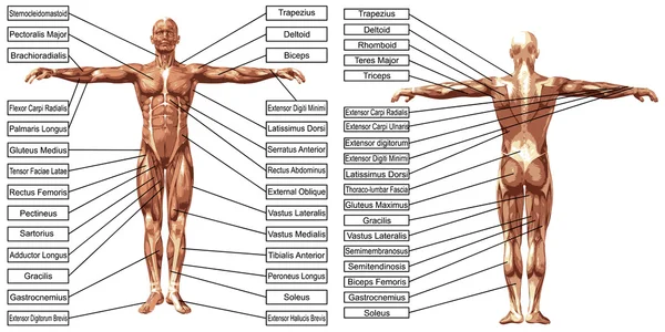
Back Muscles Diagram Labeled / Vector 3d Man Muscles Anatomy With Text Isolated On White Background 126591818 Larastock - The former two groups, superficial and intermediate, are referred to as the extrinsic back muscles.
byAdmin•
0
Back Muscles Diagram Labeled / Vector 3d Man Muscles Anatomy With Text Isolated On White Background 126591818 Larastock - The former two groups, superficial and intermediate, are referred to as the extrinsic back muscles.. Anatomynote.com found anatomy of back muscles diagram from plenty of anatomical pictures on the internet. They also attach your shoulders and pelvis to the trunk, creating a bridge between. Muscles make up a large part of the anatomy (structure) of the back. They start at the top of the neck and go down to the tailbone. There are three major groups of back muscles:.
Lower back muscle and hip pain may also be caused by stenosis in the spine. We hope this picture anatomy of back muscles diagram can help you study and research. Within this group of back muscles you will find the latissimus dorsi, the trapezius, levator scapulae and the rhomboids. This picture also contains humerus, olecranon process of ulna, deep to tendon and so on. Your back consists of three distinct layers of muscles, namely the superficial layer, the intermediate layer, and the deep layer.
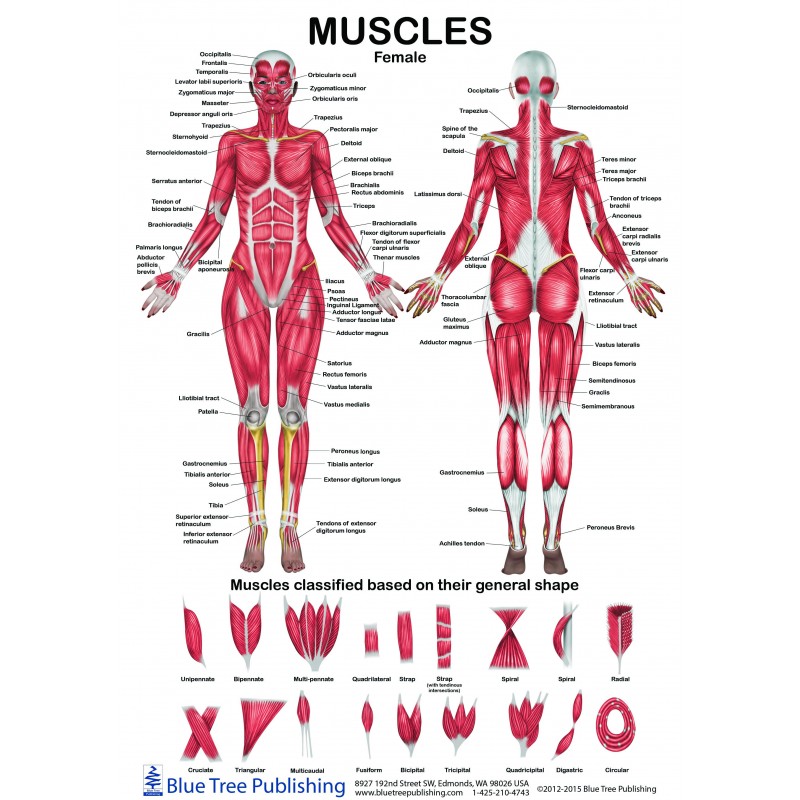
Female Male Muscle Anatomical Chart from www.bluetreepublishing.com To learn more about the anatomy of the spine, watch this video. This labeled human muscular system chart illustrates the major muscle groups in the back (posterior) view and the front (anterior) view. Claim your free copy of the client back care guide today. Reproductive · june 6, 2021. The fibres attach to the clavicle, acromion and the scapula spine. Anterior rami of upper thoracic the deep or intrinsic muscles of the back extend from the pelvis to the skull and are innervated by segmental. The muscles that make up the quadriceps are the strongest and leanest of all muscles in the body. Human anatomy diagrams show internal organs, cells, systems, conditions, symptoms and sickness information and/or tips for healthy living.
Your back consists of three distinct layers of muscles, namely the superficial layer, the intermediate layer, and the deep layer.
This labeled human muscular system chart illustrates the major muscle groups in the back (posterior) view and the front (anterior) view. How many ligaments are shown in the labeled diagram. Still, many individuals pay far too little attention to them. Link to client back care guide This article will focus on the superficial group. Major muscles back muscles shoulder muscles supraspinatus muscle back workout routine sternocleidomastoid muscle muscle diagram body diagram latissimus dorsi. The muscles of the arm anatomical chart does an exemplary job of examining the individual muscles that make up this area of the human body, and how they work together in processes such as motion and flexibility. Claim your free copy of the client back care guide today. To learn more about the anatomy of the spine, watch this video. Upper back anatomy muscles anatomy drawing diagram. The muscles of the back can be divided in three main groups according to their anatomical position and function. Muscles make up a large part of the anatomy (structure) of the back. The largest muscle masses in the leg are present in the thigh and the calf.
Lower back muscle and hip pain may also be caused by stenosis in the spine. It is the most superficial of all the back muscles. Human anatomy · june 5, 2021. Broadly considered, human muscle—like the muscles of all vertebrates—is often divided into striated muscle, smooth muscle, and cardiac muscle. Lower back muscle diagram anatomy does degenerative disc disease affect the lower back muscle?
Vector 3d Man Muscles Anatomy With Text Isolated On White Background 126591818 Larastock from st2.depositphotos.com Extrinsic and intrinsic.the back functions are many, such as to house and protect the spinal cord, hold the body and head upright, and adjust the movements of the upper and lower limbs. This labeled human muscular system chart illustrates the major muscle groups in the back (posterior) view and the front (anterior) view. Reproductive · june 6, 2021. This diagram depicts diagram back muscles. It also covers some common conditions and injuries that can affect the back. Claim your free copy of the client back care guide today. Support and protect your spine; You maintain the position of the core while moving the other parts of the body..
The muscles on each side form a trapezoid shape.
12 photos of the back muscle diagrams labeled. The back muscles enable you to stand up straight; The muscles of the lower back help stabilize, rotate, flex, and extend the spinal column, which is a bony tower of 24 vertebrae that gives the body structure and houses the spinal cord. Claim your free copy of the client back care guide today. This picture also contains humerus, olecranon process of ulna, deep to tendon and so on. Link to client back care guide Attached to the vertebral column; For more anatomy content please follow us and visit our website: The fibres attach to the clavicle, acromion and the scapula spine. Upper back anatomy muscles anatomy drawing diagram. Human muscle system, the muscles of the human body that work the skeletal system, that are under voluntary control, and that are concerned with movement, posture, and balance. Superficial back muscles, intermediate back muscles and intrinsic back muscles.the intrinsic muscles are named as such because their embryological development begins in the back, oppose to the superficial and intermediate back muscles which develop elsewhere and are therefore classed as extrinsic muscles. Attached to the shoulder girdle intermediate:
This is a diagram of the larger and more surface muscles of the low back. It comprises the vertebral column (spine) and two compartments of back muscles; Link to client back care guide The muscles of the back. Key muscles of the hip :
Labeled Anatomy Chart Male Back Muscles Stock Illustration 1423709246 from image.shutterstock.com The trapezius is a broad, flat and triangular muscle. Muscles found in the deep group include the spinotransversales, erector spinae (composed of the iliocostalis, longissimus, and spinalis), the transversospinales, and the segmental muscles. They start at the top of the neck and go down to the tailbone. Link to client back care guide This article gives an overview of the back's structure and its major muscles. Human muscle system, the muscles of the human body that work the skeletal system, that are under voluntary control, and that are concerned with movement, posture, and balance. This labeled human muscular system chart illustrates the major muscle groups in the back (posterior) view and the front (anterior) view. They also attach your shoulders and pelvis to the trunk, creating a bridge between.
Piriformis muscle anatomy ultrasound 12 photos of the piriformis muscle anatomy ultrasound piriformis muscle anatomy ultrasound, human muscles, piriformis muscle anatomy ultrasound.
The back muscles enable you to stand up straight; It also covers some common conditions and injuries that can affect the back. Extrinsic and intrinsic.the back functions are many, such as to house and protect the spinal cord, hold the body and head upright, and adjust the movements of the upper and lower limbs. Lower back muscle and hip pain may also be caused by stenosis in the spine. The muscles of the back can be arranged into 3 categories based on their location: Anatomynote.com found anatomy of back muscles diagram from plenty of anatomical pictures on the internet. This article gives an overview of the back's structure and its major muscles. Another common cause of lower back and hip pain is disc injury. The trapezius is a broad, flat and triangular muscle. Related posts of muscles of the lower back and buttocks diagram piriformis muscle anatomy ultrasound. And reach, pull and extend your arms and torso. Support and protect your spine; Piriformis muscle anatomy ultrasound 12 photos of the piriformis muscle anatomy ultrasound piriformis muscle anatomy ultrasound, human muscles, piriformis muscle anatomy ultrasound.
Claim your free copy of the client back care guide today back muscles diagram. Extrinsic and intrinsic.the back functions are many, such as to house and protect the spinal cord, hold the body and head upright, and adjust the movements of the upper and lower limbs.